Quiz Case #4

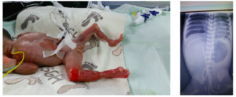
This is a premature baby girl born at 27 weeks gestation, with a birth weight of 620 g. Her length and occipital frontal circumference are appropriate for age. Her parents are non-consanguineous and healthy. Antenatal history was unremarkable. She presents at birth with extensive areas of raw denuded skin over both ears, upper limbs, lower limbs and around the umbilicus. She also had hypoplastic ears bilaterally with hypoplastic left lower limb (Figure A). The 2D echocardiogram showed a patent ductus arteriosus and small patent foramen ovale, with a structurally normal heart. A chest and abdominal radiograph done to check the position of her umbilical lines was found to be abnormal (Figure B).
This is a premature baby girl born at 27 weeks gestation, with a birth weight of 620 g. Her length and occipital frontal circumference are appropriate for age. Her parents are non-consanguineous and healthy. Antenatal history was unremarkable. She presents at birth with extensive areas of raw denuded skin over both ears, upper limbs, lower limbs and around the umbilicus. She also had hypoplastic ears bilaterally with hypoplastic left lower limb (Figure A). The 2D echocardiogram showed a patent ductus arteriosus and small patent foramen ovale, with a structurally normal heart. A chest and abdominal radiograph done to check the position of her umbilical lines was found to be abnormal (Figure B).
This child has JEBPA with extensive scarring in utero, which resulted in hypoplasia of her ears and left lower limb. Blood culture and bacterial swab culture from the denuded skin were negative. Mutation analysis revealed a compound heterozygous mutation for the c.912C>G (p.TYR304*) (from her father) and c.2251C>T (p.Arg751*) (from her mother) variants in the ITGB4 gene. Immunofluorescent mapping of a fresh blister also showed absent staining to Integrin β-4, a protein that forms integrin which regulates skin integrity.
Discussion
The radiograph shows a dilated gastric bubble with paucity of gas in the distal bowels.
It is clinically difficult to clinically differentiate between junctional epidermolysis bullosa and recessive dystrophic epidermolysis bullosa at birth without molecular genetic testing. The presence of pyloric atresia would make JEBPA the most likely diagnosis.
Staphylococcal scalded skin syndrome can cause extensive desquamation of the skin and could be fatal in a premature infant. However, the bacterial swab was negative.
Intrauterine HSV disease may present with a triad of clinical findings including cutaneous (scaring, active vesicles, dyspigmentation and or aplasia cutis), ophthalmologic (retinal dysplasia, optic atrophy and chorioretinitis) and neurological (microcephaly, hydraencephaly and or intracranial calcifications) involvement. . The large areas of skin denudation is not typical of HSV disease in this case.
The possibility of Trisomy 18 may be to be detected on antenatal screening. Clinically, the baby did not have other dysmorphic features of Trisomy 18 such as rocker bottom feet.
Congenital epidermolysis bullosa
Inherited epidermolysis bullosa (EB) is a rare genodermatosis characterised by mechanical fragility of epithelial tissues in the skin and mucous membranes, resulting in recurrent blistering and erosions which heal poorly. There are four major groups based on the ultrastructural level within which blisters develop in the affected tissues (EB simplex, junctional EB (JEB), dominant dystrophic EB and recessive dystrophic EB).
Genetic testing is done to confirm mutation of the affected gene in congenital EB. The diagnosis is also supported by a skin punch biopsy of the edge of a new blister for immunofluorescent mapping to identify the missing protein in the basement membrane.
JEB is characterized by an autosomal recessive inherited pattern. A rare subtype of JEB with pyloric atresia (JEB-PA) presents with gastrointestinal, genitourinary and respiratory epithelial lesions, and involves widespread blistering and erosions of the integument and oral mucosa. It is associated with mutations in ITGB4 (Integrin -4), or less commonly ITGA6 (Integrin 6) which encode the beta4 (b4) or alpha6 (a6) subunit of integrin, respectively. Integrin a6b4 is involved in the adhesion of the basal keratinocytes to the underlying mesenchyme and plays an important role in maintaining the integrity of skin and epithelial linings. Mutation in the plectin (PLEC1) gene has been recently found to also be associated with JEB-PA.
These patients will require multidisciplinary team care including the neonatologist, paediatric dermatologist, paediatric surgeons, wound nurse and other paediatric subspecialties depending on which other mucosal sites are affected.
Life expectancy of children with JEB is poor and about half do not survive past the first year of life while most die before 5 years old. The prognosis was very grim for this case given her extreme prematurity, pyloric atresia and fragile skin that made it difficult to secure her umbilical lines for nutrition and treatment. She expired at two weeks old despite intensive care support.
References:
- Fine, J.D., et al., Inherited epidermolysis bullosa: updated recommendations on diagnosis and classification. J Am Acad Dermatol, 2014. 70(6): p. 1103-26.
- Chung, H.J. and J. Uitto, Epidermolysis bullosa with pyloric atresia. Dermatol Clin, 2010. 28(1): p. 43-54.
Author:
Dr Lynette Wee Wei Yi
MBBS, MRCPCH, MMED (Paeds)
Consultant, Paediatric dermatology
Dermatology service
KK Women’s and Children’s Hospital