


?
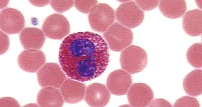
Eosinophils
Bilobed
Red cytoplasm
Blue nucleus
Grannulocytes
Staining Giemsa
Blood- Eosinophils - staining Giemsa


?
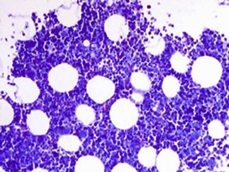
Bone Marrow
Staining Giemsa
Adipocytes
Stroma
Megakaryocytes
Hemapoietic cells
Sinusoids
Nucleus of endothelial cells
Arterioles
Endothelial cells
Smooth muscle cells
Bone marrow staining- giemsa

?
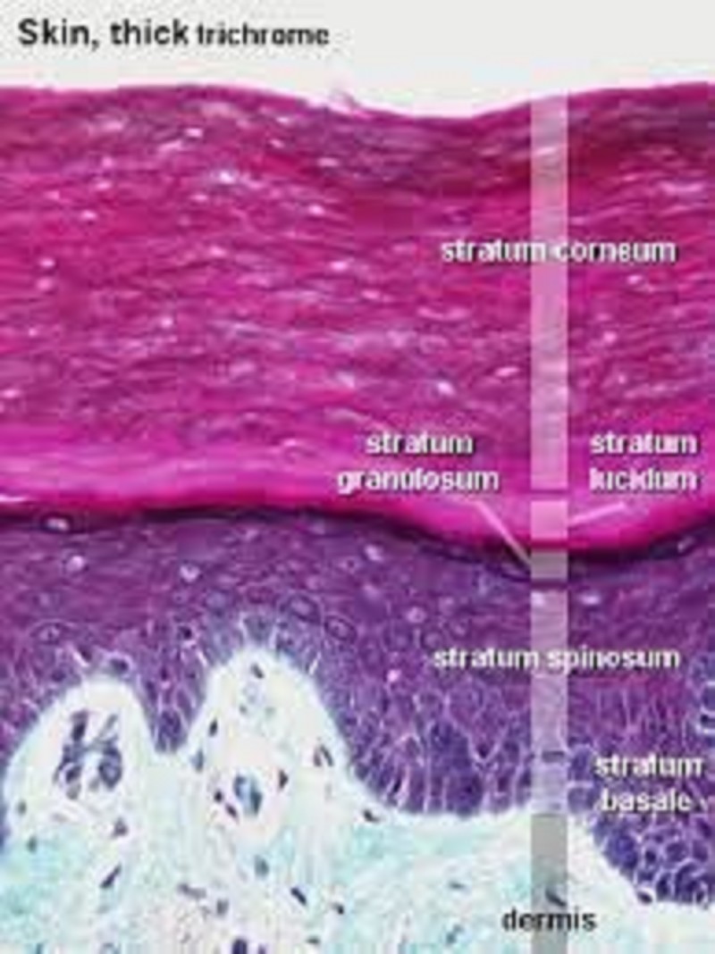
Epidermis of thick skin
Stratum basale
Stratum spinosum
Stratum granulosum- Granules
Stratum lucidum- No cells
Stratum corneum - Dead keratinocytes
Dermal papillae = the connection of the epidermis to the skin
Epidermal ridges
Staining H&E
Epidermis of thick skin- staining H&E
?
0%
0

0%
0
0%
0
0%
0
0%
0
0%
0
0%
0


?
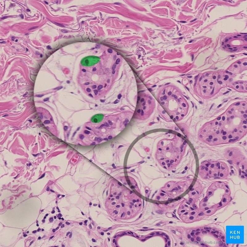
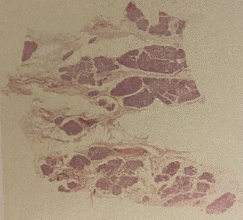
Mammary gland
Staining H&E
Lobes divided within each other
Lactiferous sinus with septa
Blood vessels inside CT septa
Excretory ducts
Mammary gland - Staining H&E
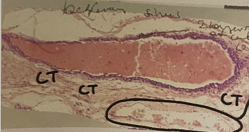
?
Double layer of cuboidal cells
Active
Simple squamous epithelium, with erythrocytes and blood vessels inside (within the circle)
Connective Tissue
Lactiferous sinus within septa- staining H&E
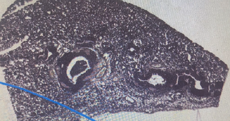
?
Lungs
Staining H&E
Respiratory Epithelium - Psudostratified columnar ciliated, has glands
Bronchus
Bronchioles
Alveoli
Lungs - staining H&E
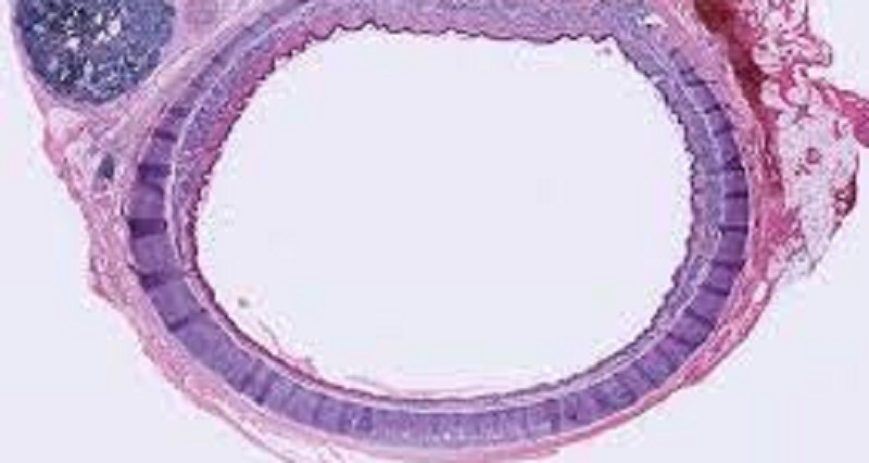
?
Trachea
Staining H&E
Regular Lumen
Adipose tissue around trachea
Mucosa
Submucosa
Adventina
Trachea - staining H&E
?
0%
0
0%
0
0%
0
0%
0
0%
0
0%
0


?
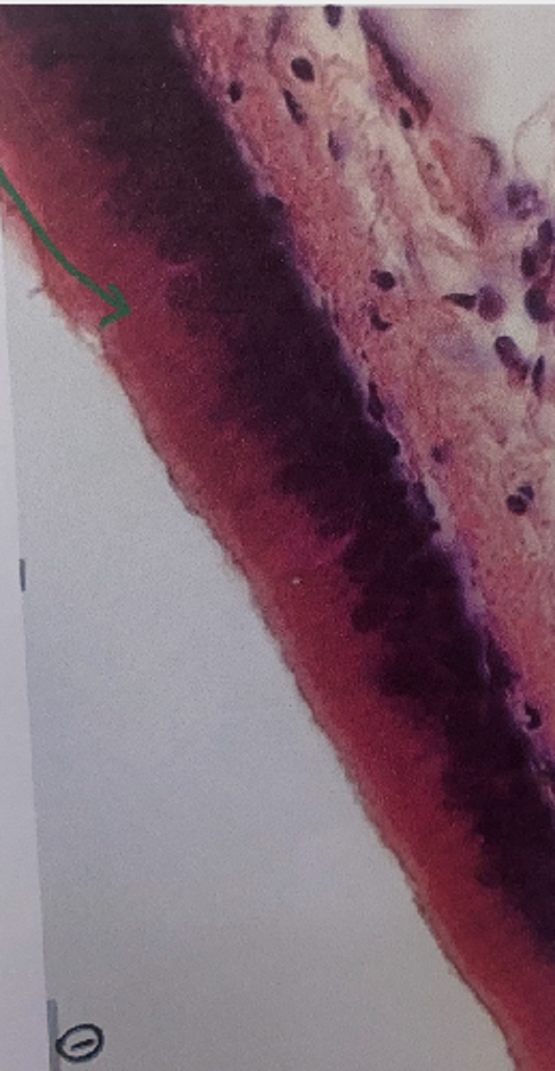
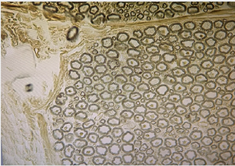
Staining- Resorcinfuchsin
Tunica intima- Endothelium + subendothelial layer
Tunica Media- elastic lamina, fenestrated, smooth muscle cells (vascularized)
Tunica adventina- Loose CT, adipose tissue, nerves, vasa Vasorum
Elastic artery - staining Resorcinfuchsin, (details)

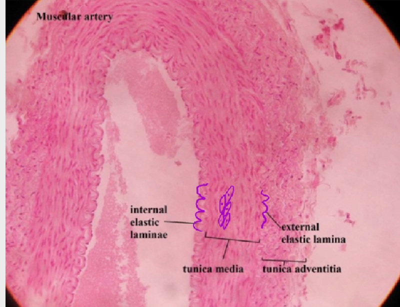
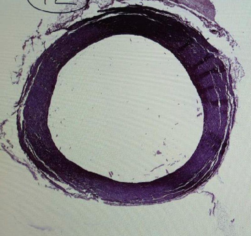
?
Tunica intima- prominat internal elastic lamina, visible endothelial cells
Tunica media- Smooth muscle cells, collagen bundles, a lot of EMC, External elastic lamina
Tunica Adventitia- visible CT, nerves, vasa vasorum
Erythocytes in lumen
Muscular artery - staining H&E
?
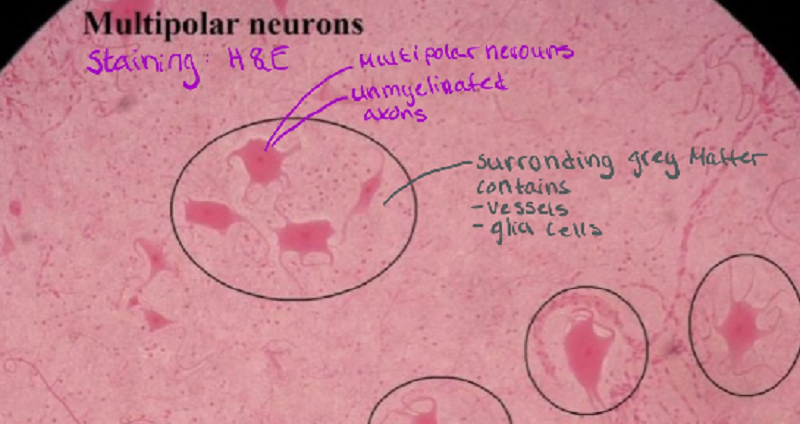
Perikaryon/ body
Nuclei and nucleous
Dendrites/ processes
Nissls bodies
Glia cells
Multipolar Neurons - staining H&E
?
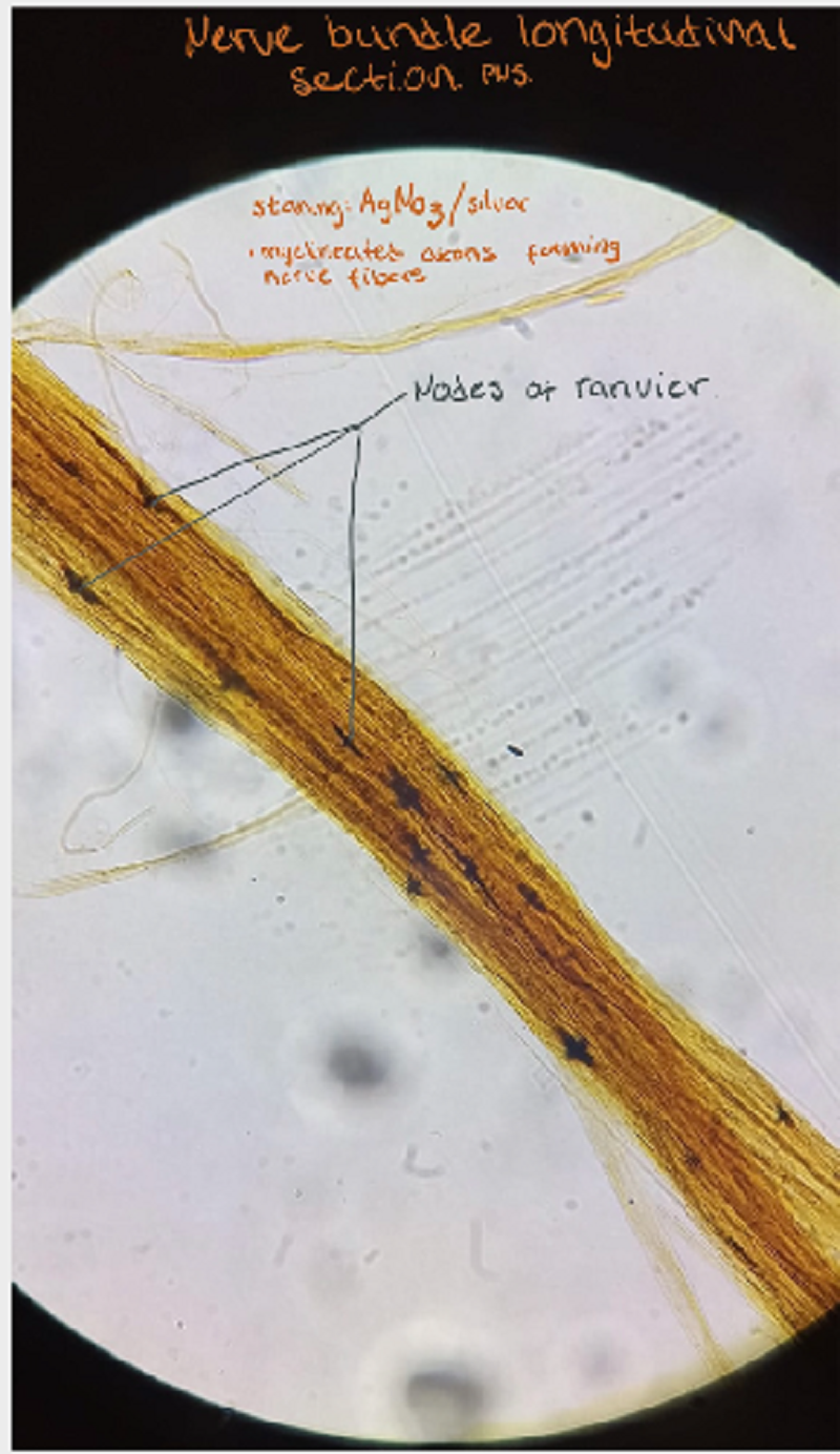
Axons, strings, process of perikaryon, covered by myelin sheat
Nodes of Ranvier
Nerve bundles in PNS - Staining AgNo3/silver
?
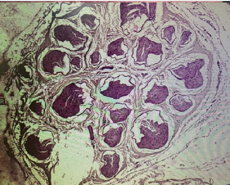
Nerve bundles cross section
Staining H&E
Epineurium
Perineurium
Endoneurium
Nerve bundles cross section- staining H&E
?
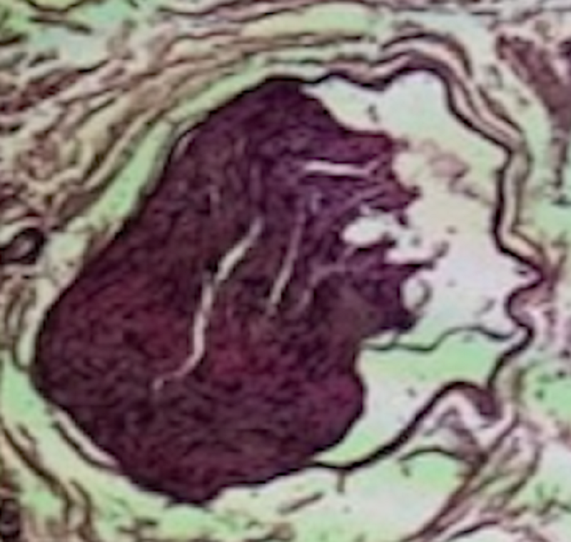
Epineurium- Dens CT, surround whole nerve
Perineurium- Dense CT, surrounds nerve bundles
Endoneurium- Loose CT, surrounds each fiber, schwann cells, fibroblast in CT
Nerve bundles cross section- H&E staining

?
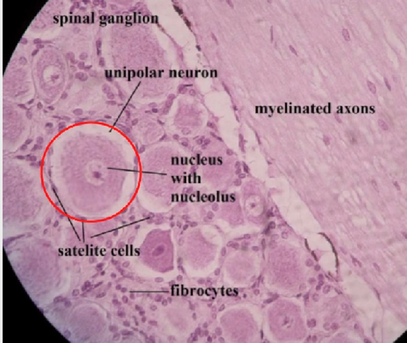
Surrounded by CT capsule
Perikaryons og psudounipolar neuron
Nucleus + nucleous
Satellite cells
Process of neurons
Nissl bodies
Longitudinal axons- nuclei of schwann cells, Axons +myelin sheat
Perineurium- covers bundles
Spinal Ganglia- staining H&E
?
Myelin sheats- contains axons inside
Endoneurium around the axons- loose CT
Perineurium- surrounds nerve bundles, Dens CT
Nerve Bundles cross section- OsO4
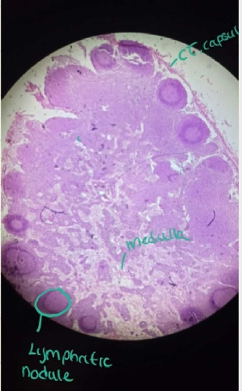
?
Surrounding CT capsule
Subcapsular sinus
HEVs
Cortex
Paracortex
Medulla
Blood vessels in every layer
Lymph Nodes- stainig H&E
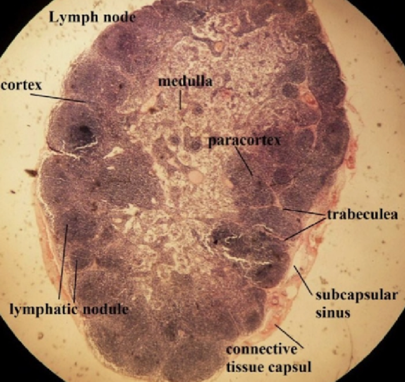
?
Subcapsular sinus
HEV´s- between lymphatic nodules
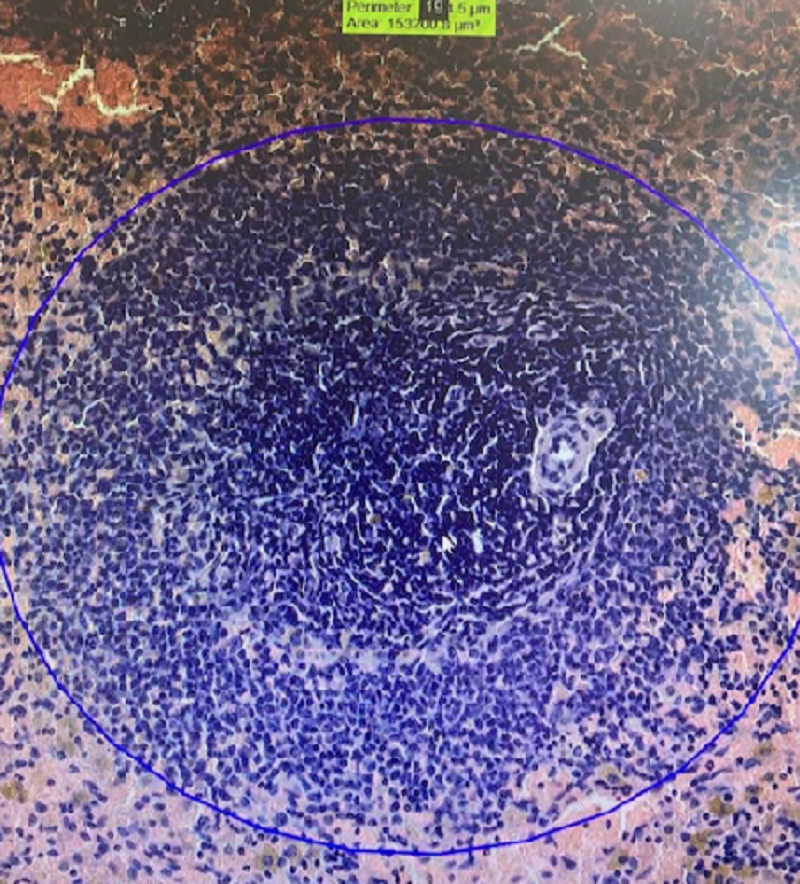
Cortex- lymphatic nodules
Paracortex- active nodules, Trabecular sinus, lymphatic nodules
Medulla- medullary sinus, medullary cords
Blood vessels in every layer
Lymphe nodes, staining H&E
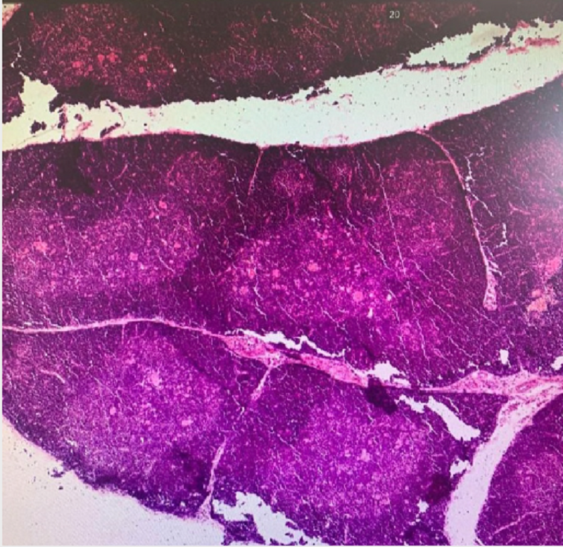
?
Surrounded by CT capsule
Trabeculi w/ B.V in sinusoids
Trabeculi w/smooth muscle cell inside B.V
Divided into white- & red- pulp
Blood vessels
Spleen - staining H&E
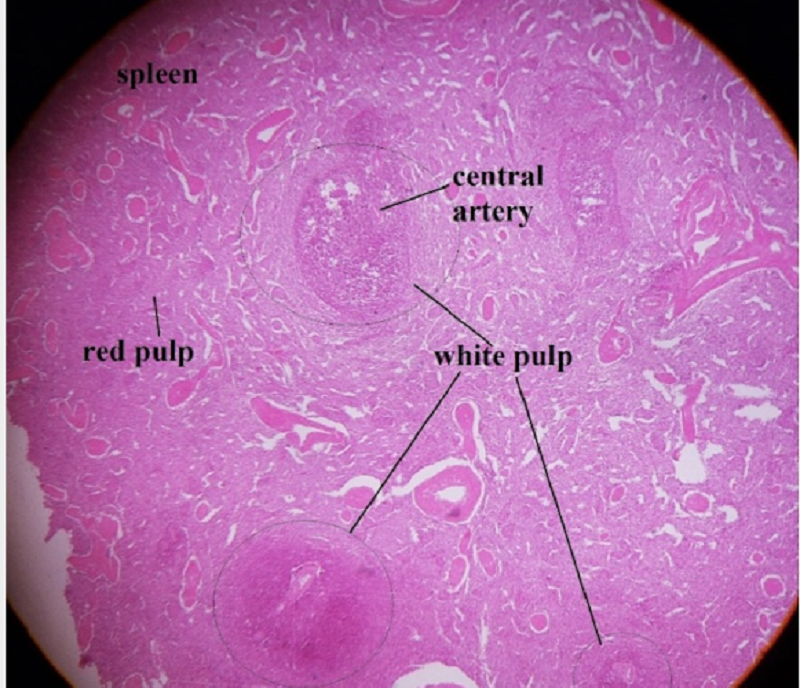
?
Trabeculi w/ B.V in sinusoids
Trabeculi w/smooth muscle cell inside B.V
White pulp- Lymphoid nodules, central artery, PALS, t-lymphocytes
Red pulp- macrophages, plasma cells, T+B cells, reticular fibers
Spleen - staining H&E

?
Surrounded by CT capsule
Cortex- pre-T lymphocytes
Medulla- T-lymphocytes, Hassal corpusle
Stroma of thymus- T-cells, epithelial cells, hassal bodies
Thymus- staining H&E

{"name":"?", "url":"https://www.quiz-maker.com/QPREVIEW","txt":"?, ?, ?","img":"https://www.quiz-maker.com/3012/CDN/73-3366948/blood.png?sz=1200"}