Dental Imagery II 2

Dental Imagery II Quiz
Welcome to the Dental Imagery II Quiz! This informative quiz is designed for dental professionals and students to test their knowledge on various concepts related to dental imaging, oral assessments, and dental conditions.
Prepare to challenge yourself with thought-provoking questions that cover:
- Radiographic techniques
- Dental anomalies
- Bone structure and pathology
- Interpretation of dental images
Many techniques are available to the dentist for carrying out oral assessments. Which of the following is only used to determine if a tooth is dead?
Hot stimulus
Radiograph
Study models
Trans illumination
A patient come to the dental clinic and is diagnosed with dental anodontia. Which one of the following features does the patient exhibit?
Congenitally absent teeth
Enlarged teeth
Microdontia
Supernumerary teeth
. Common radiographic projection for fracture the ramus of mandible?
Periapical
OPG
Bitwing
Arthroscopy
Common radiographic projection for fracture the canine region?
0º (OM)
30º (OM)
OPG or oblique lateral
Periapical
Common radiographic projection for investigation site of fracture of dento-alveolar?
Periapical
Bit wing
Arthroscopy
Panoramic
In case the fracture displacement to overlap of the fragment is shown in radiograph:?
Radiopaque line
Radiolucency line
No evident
Radiolucency total
In case fracture step deformation due to separation and displacement of fragment is shown in radiograph:?
Radiopaque line
Radiolucency line
No evident
Radiolucency total
In case fracture no displacement of the fragment is shown in radiograph:?
Radiolucency line
Radiopaque line
No evident
Radiolucency total
Main radiographic features of large squamous cell carcinoma radiographic investigation shown:?
Radiopaque total
Radiolucency total
Radiolucency total
Periapical
Which methods are used commonly to access tooth deep of third molar?
Winter’s line
Radiolucency total
Loss of the tramline
Parallax plan
What is the fusion teeth?
Two teeth joined together from the fusion of adjacent tooth germs
Two teeth joined together but arising from a single tooth germ
Two teeth joined together by cementum
Large teeth
What is the microdontia?
Two teeth joined together but arising from a single tooth germ
Two teeth joined together by cementum
Small teeth, including rudimentary teeth
Two teeth joined together from the fusion of adjacent tooth germs
Which one is the other mandibular anomalies?
Condylar hypoplasia
Parallax plan
Compound odontoma
Concrescence
Which technique that easy to interpretation of unerupted maxillary canines?
Parallax in the horizontal plan
Vertex occlusal
Periapical
Panoramic
Which essential requirements for interpretation?
Viewing box
The patient
Exposure factor and film density
Viewing box, magnifying glass, SDI X-ray reader
What the meaning of radiolucent in the radiograph?
Black color
White color
Yellow with red color
Gray color
Which is the good place for film interpretation?
Darkness viewing room with viewing box
Bright room
Normal room
Under sunlight
. For X-ray interpretation, what is the knowledge for?
Anatomical structure of teeth and skull
The type of radiograph
Panoramic radiograph
Systemic approach
For X-ray interpretation, what is the knowledge for?
Pathological and specific lesson
The patient
Sign of inflammatory
Exposure factor of film
. Which methods are used commonly to assess tooth depth?
Loss of tramline
White line
Narrowing of tramline
Using the root of the second molar as a guide
Which line that draw from cervical first molar to second molar?
Red line
White line
Amber line
Winter’s line
. Which line that draw from occlusal surface of the first molar to second molar?
Red line
Amber line
Winter’s line
White line
When is easy time of using the root of mandibular second molar as guide?
Coronal third
Apical third
Middle third
Over 5 mm
complex odontoma is:?
Small tooth
Large tooth
A lot of teeth that mixed together
A lot of teeth that stay together
Compound odontoma is:?
Small tooth
Large tooth
A lot of teeth that mixed together
A lot of teeth that stay together
What is hyperdontia?
Supernumerary teeth and supplemental teeth
Amelogenesis imperfect
Dentinal dysplasia
Turner teeth
Transposition tooth is:?
Two teeth occupying exchanged position
Complex mass of disorganized dental tissue
Made up of one or more small tooth like denticles
Two teeth joined together
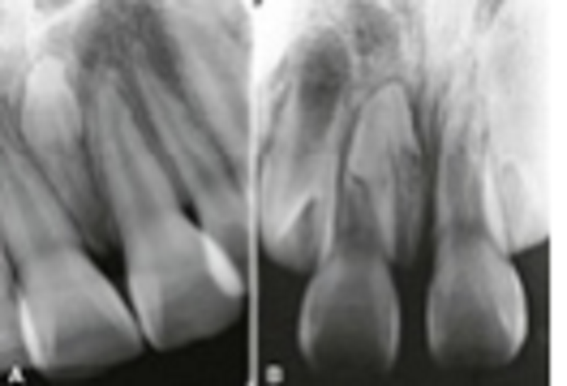
Please select the correct answer at 2-part A and B:?
Supernumerary
Supplemental
Microdontia
Shell Teeth
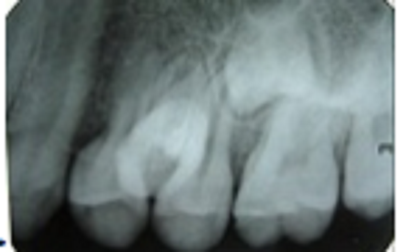
Please choose the correct answer at the arrow point:?
Supernumerary
Multiple supplemental premolar
Microdontia
Shell Teeth
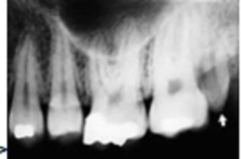
Please choose the correct answer at the arrow point:?
Supernumerary
Supplemental
Microdontia
Shell Teeth

Please choose the correct answer at the arrow point:?
Supernumerary
Supplemental
Microdontia
Shell Teeth
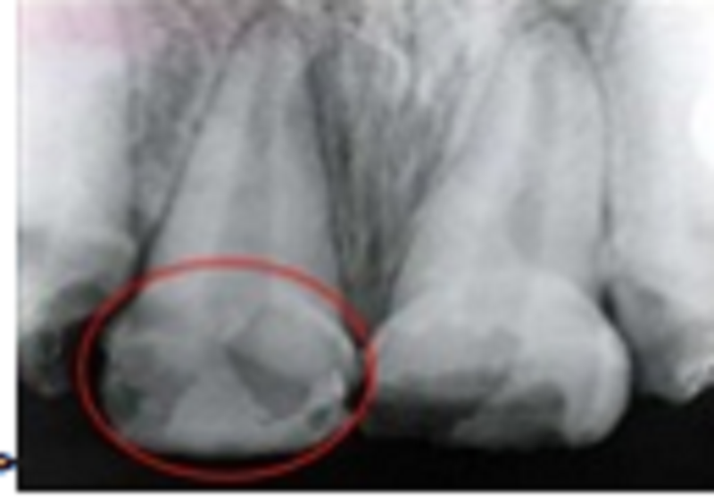
Please choose the correct answer at the red round line:?
Supernumerary
Supplemental
Microdontia
Enamel defects of amelogenesis imperfecta
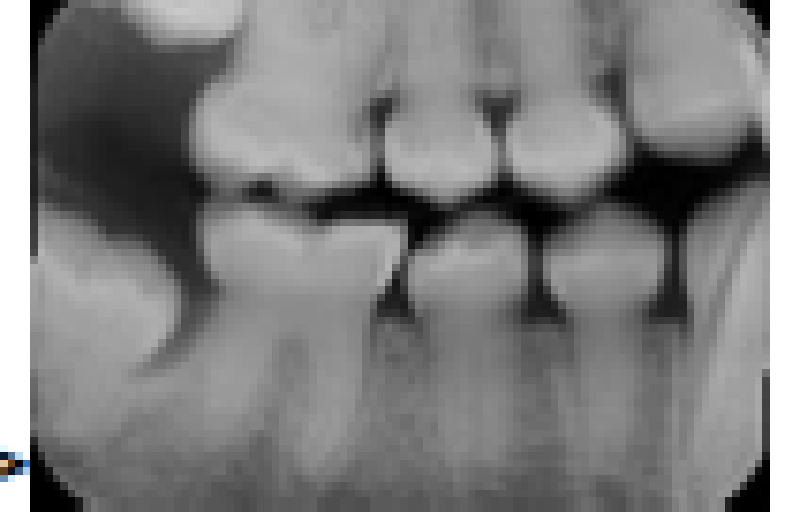
Please choose the correct answer at the film:?
Supernumerary
Defect of dentinogenesis imperfecta
Microdontia
Enamel defects of amelogenesis imperfecta
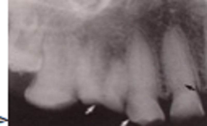
What kind of film in this Radiograph?
Periapical of maxillary premolars and molars
Periapical of mandibular premolars and molars
Anoramic of maxillary premolars and molars
Bitewing of maxillary
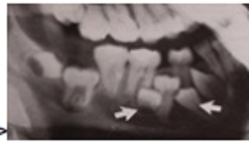
What kind of film in this Radiograph?
Periapical of maxillary premolars and molars
Periapical of mandibular premolars and molars
Panoramic of maxillary premolars and molars
Oblique Lateral
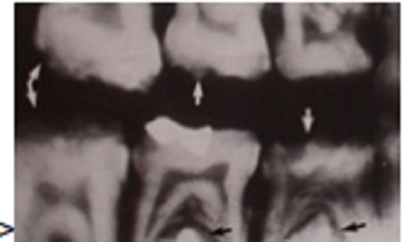
What kind of film in this Radiograph?
Periapical of maxillary premolars and molars
Periapical of mandibular premolars and molars
Panoramic of maxillary premolars and molars
Bitewing
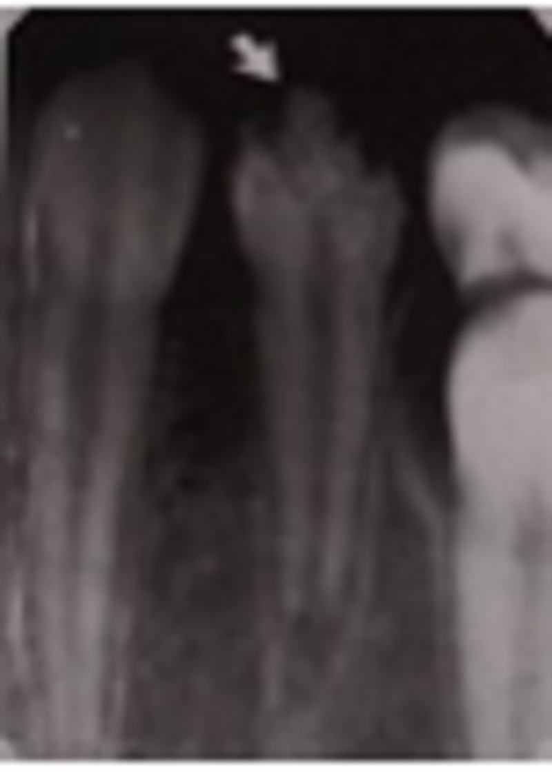
Please choose the correct answer at the arrow point:?
Supernumerary
Supplemental
Enamel defects of amelogenesis imperfecta
Enamel defects of a Turner tooth
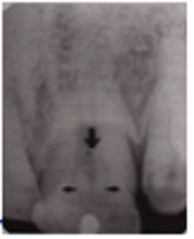
Please choose the correct answer at the arrow point:?
Supernumerary
Defect of dentinogenesis imperfecta
Microdontia
Fusion of tooth
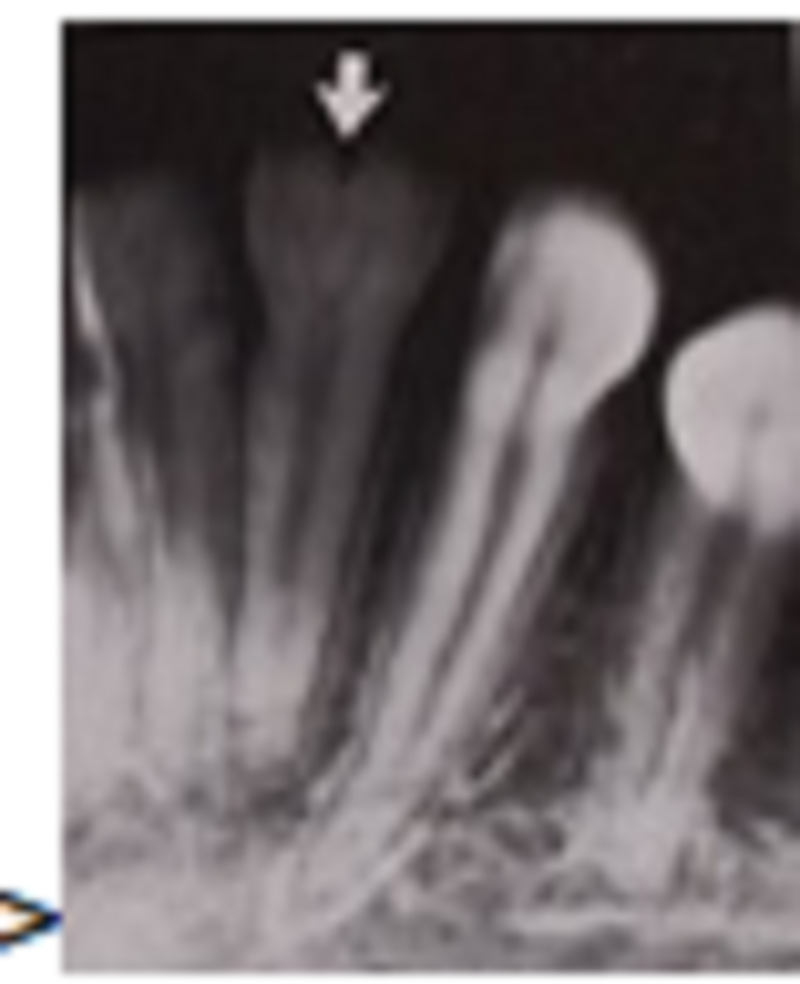
Please choose the correct answer at the arrow point:?
Supernumerary
Gemination of tooth
Caries of tooth
Fusion of tooth
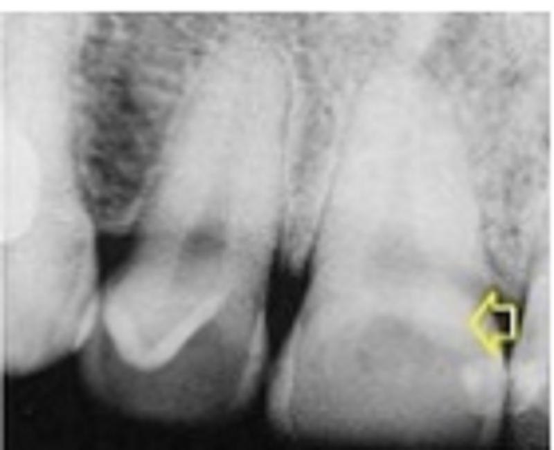
Please choose the correct answer at the arrow point:?
Macrodontia
Gemination of tooth
Caries of tooth
Fusion of toot
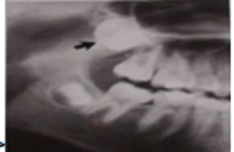
What kind of radiograph?
Part of a dental panoramic tomography
Bitewing radiograph
Periapical
Oblique lateral

Please choose the correct answer at the arrow point:?
Macrodontia
Dens-in-dente
Caries of tooth
Fusion of tooth
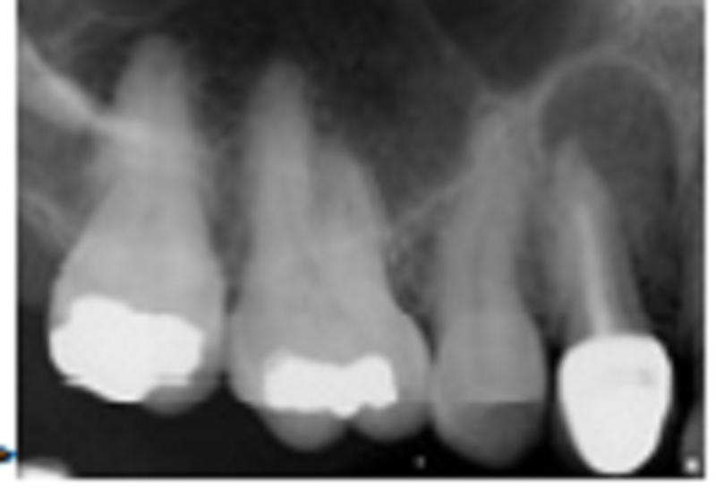
What happened in the root of tooth #4?
Good Bone
Periapical / radicular cyst
Fusion of tooth
Over exposure of radiograph
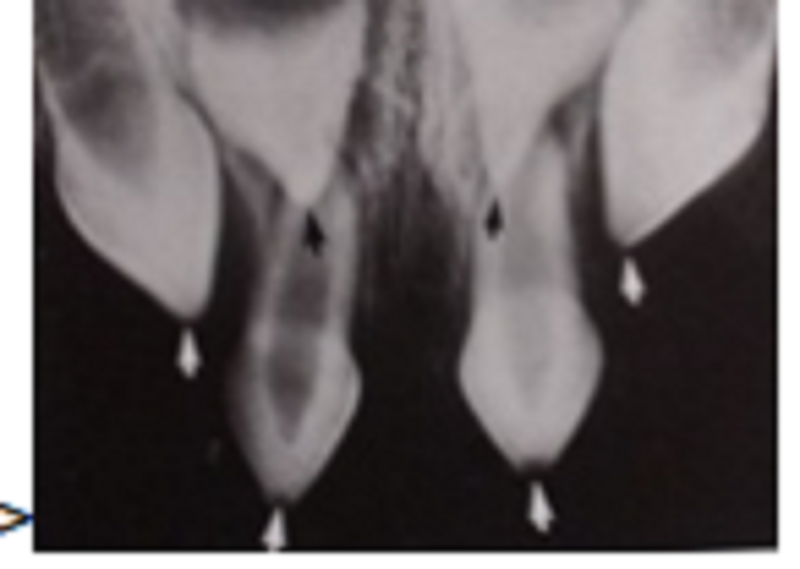
Please choose the correct answer at the arrow point:?
Dens-in-dente
Microdontia
Ectodermal dysplasia
Supernumerary
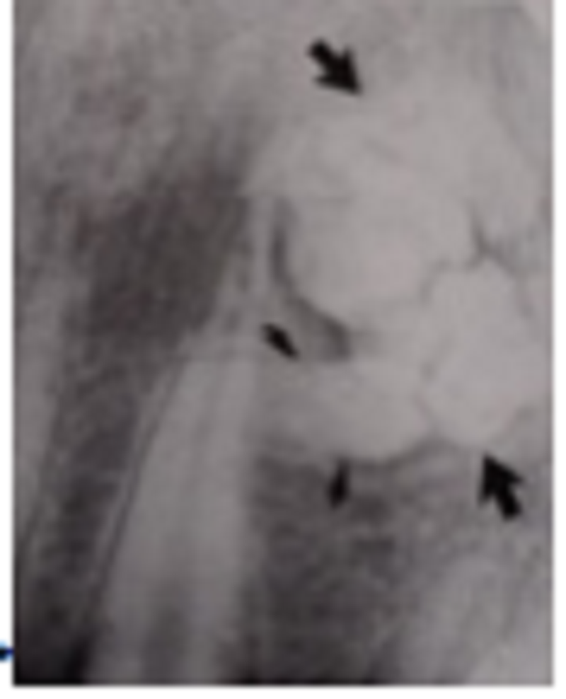
Please choose the correct answer at the arrow point:?
Dens-in-dente
Compound odontoma in the anterior maxilla
Microdontia
Complex odontoma
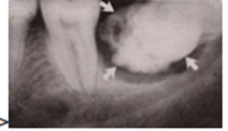
Please choose the correct answer at the arrow point:?
Dens-in-dente
Compound odontoma in the posterior mandibula
Microdontia
Complex odontoma
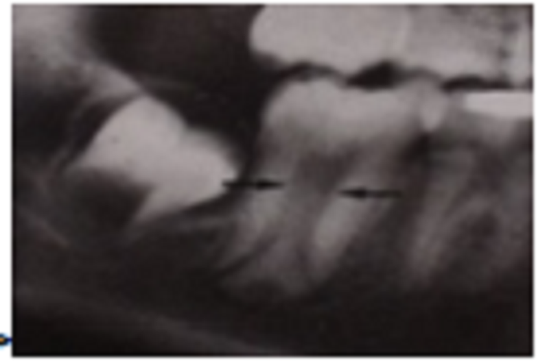
Please choose the correct answer at the arrow point:?
Dens-in-dente
Taurodontism
Microdontia
Complex odontoma
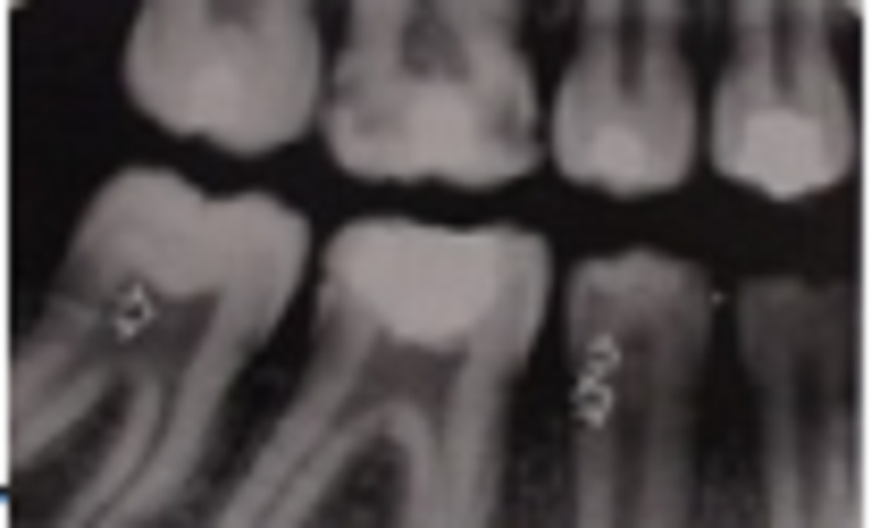
Please choose the correct answer from arrow point at radiopaque zone:?
Pulpstone
Taurodontism
Microdontia
Complex odontoma
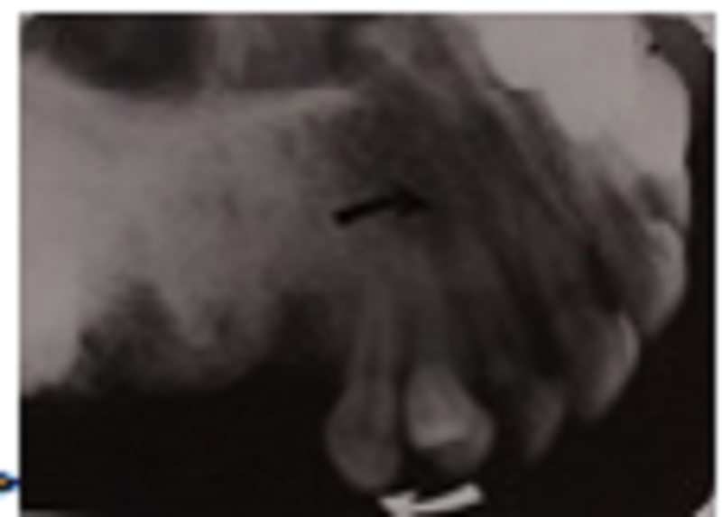
Please choose the correct answer at the arrow point:?
Pulpstone
Taurodontism
Transposition of tooth
Complex odontoma
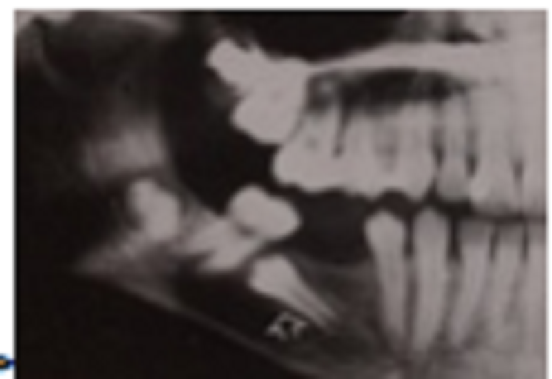
Please choose the correct answer at the arrow point:?
Pulpstone
Wandering tooth
Microdontia
Complex odontoma
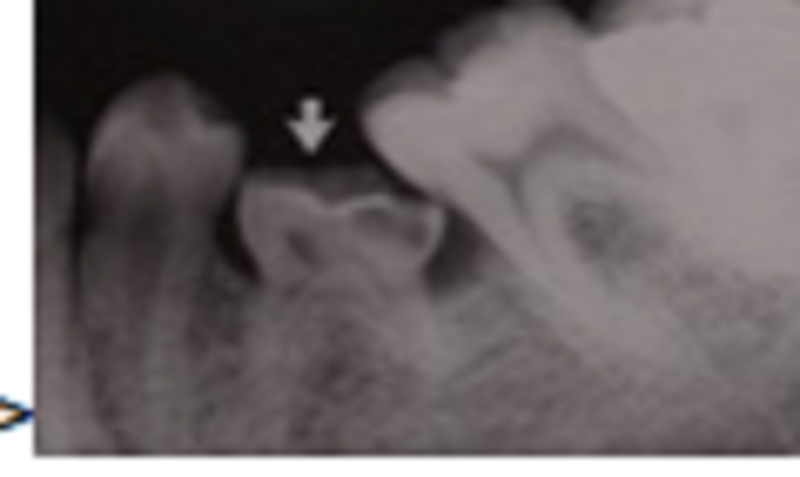
Please choose the correct answer at the arrow point:?
Submerging or infra-occlusal
Wandering tooth
Microdontia
Complex odontoma
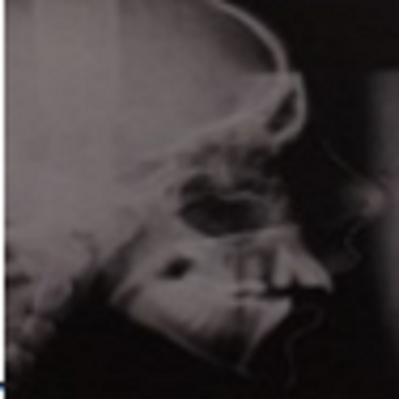
Please choose the correct answer in the radiograph:?
Submerging or infra-occlusal
Micrognathia
Classification class II
Macrognathia

Please choose the correct answer in the radiograph:?
Classification class II
Micrognathia
Classification class I
Macrognathia

Please choose the correct answer on the radiograph:?
Bifid of condyle
Cleft palate
Classification class I
Macrognathia
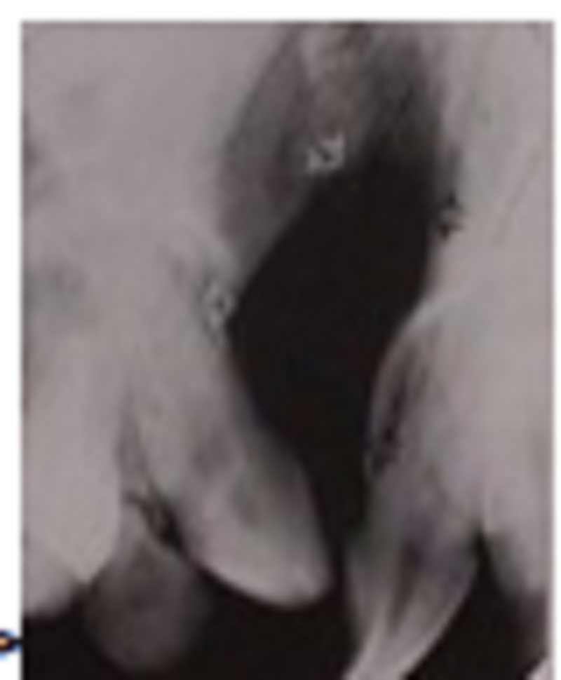
Please choose the correct answer on this radiograph:?
Bifid of condyle
Cleft palate
Classification class I
Macrognathia
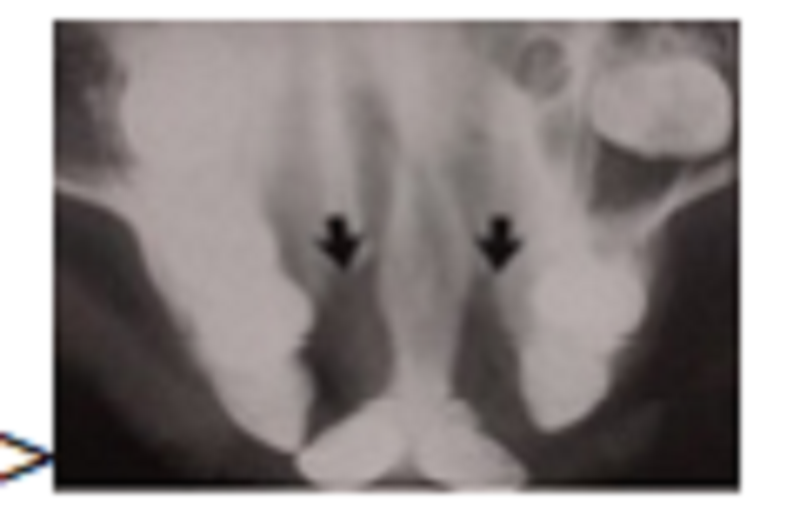
Please choose the correct answer on this radiograph:?
Bifid of condyle
Bilateral cleft palate
Classification class I
Macrognathia
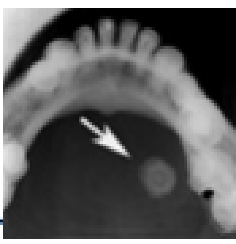
Answer in the arrow point on film x-ray:?
Long calcified stylo-hypoid ligament
Tramline
Stone in the salivary gland
ID canal
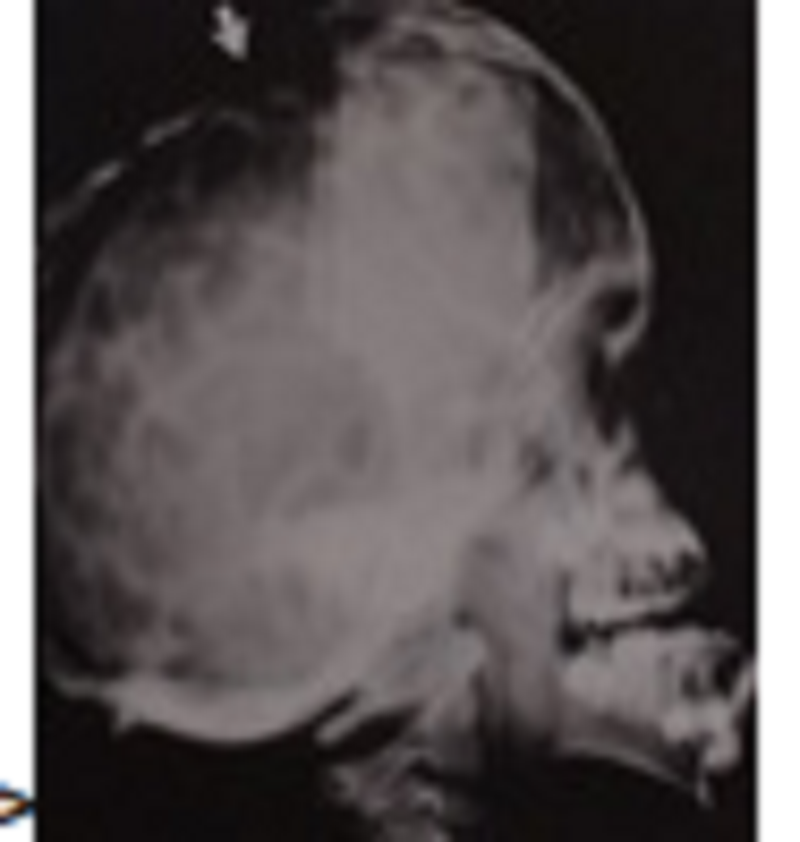
The arrow point in film x-ray, What diagnosis?
Crouzon’s and apert’s syndromes
Micrognathia
Macrognathia
Cyst of skull
{"name":"Dental Imagery II 2", "url":"https://www.supersurvey.com/QPREVIEW","txt":"Welcome to the Dental Imagery II Quiz! This informative quiz is designed for dental professionals and students to test their knowledge on various concepts related to dental imaging, oral assessments, and dental conditions.Prepare to challenge yourself with thought-provoking questions that cover:Radiographic techniquesDental anomaliesBone structure and pathologyInterpretation of dental images","img":"https:/images/course6.png"}
More Surveys
Dental Imagery II 1
723640
Complete Denture 1 សូមមេត្តាកុំចែកចាយ Link ដោយគ្មានការអនុញ្ញាត
60300
Endodontics Knowledge Quiz
7437250
Periodontology
5326660
Oral pathology 150-200
512630
Periodontology
5728819
Partial Denture 2
763825
Oral Surgery/Dr.Pin Bosara
211079
Fix Prostodontic 5
60300
Endodontics
6030306
Endodontics
6030282
Endodontic 1
723625


